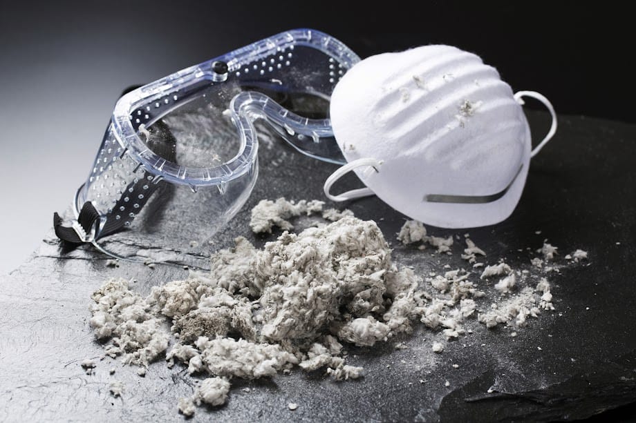
Newest Posts

What are Nucleosides, Nucleic Acid, Nucleotides

“Nucleic acid” is found within the name of DNA (Deoxyribose nucleic acid) and RNA (Ribose nucleic acid), but aren’t DNA & RNA made up of nucleotides?

NUCLEOSIDE :

is the ribose or deoxyribose sugar bound with a nitrogenous base (purine or pyrimidine) without the attachment of a phosphate group

NUCLEOTIDE

is made of:

1. Five carbon sugar also known as a pentose (pent = five) sugar. This pentose, or five carbon sugar, may either be deoxyribose or ribose. The difference between the two is at the 2nd carbon of the five ring structure. If there is NO “OH” or hydroxyl group attached to that 2nd carbon, it is called a deoxyribose. If there is an “OH” or hydroxyl group attached, it is called a ribose.

2. A tri-phosphate group attached at the 5’ carbon of the pentose

3. A base attached at the 1’ carbon atom of the pentose. This base isn’t just any base, it is a Nitrogen containing ring structure, and can be either purines or pyrimidines.

Purines (found in both RNA and DNA)

Adenine

Guanine

Pyrimidines (in DNA)

Cytosine

Thymine (not in RNA)

Uracil 
(found in RNA)

Examples of nucleotides in DNA are: dAMP, dGMP, dCMP, dTMP.

dATP isn’t considered a nucleotide because there are three phosphate groups instead of one.

Examples of nucleotides in RNA are AMP, GMP, CMP, TMP.

Note: d = deoxy (describing the sugar, ribose), AMP = adenosine mono phosphate, GMP = guanine mono phosphate, CMP = cytosine mono phosphate, TMP = thymine mono phosphate Nucleic Acid

NUCLEIC ACID

This is a sequence of nucleotides covalently bound together. The result is an alternating sugar to phosphate sequence found as the backbone of the DNA or RNA strand. Since each nucleotides are acidic, it is rational to say that a series of these nucleotides would give you an acidic polymer (nucleic acid).

Quick Review: What is the structural difference between RNA and DNA?

Viruses, Viroids, and Prions

Viruses

Viruses, viroids, and prions are all acellular pathogens. They are not within any kingdom and carry their own significant characteristics.

  1. obligate intracellular parasite – require a host to cause damage
  2. filterable – small enough to be filtrated
  3. contains an outer protein coat and inner genome
  4. has only 1 kind of nucleic acid (RNA or DNA, but never both)
  5. lacks metabolic abilities

Viruses can be found either inside a cell (intracellular) or outside of a cell (extracellular). If it is found extracellular, the virus is called a virion. A virion contains a protein coating called a capsid, which surrounds the core of the virus containing the nucleic acid (either DNA or RNA).  Together with the capsid and the DNA or RNA core is called a nucleocapsid.  Some virions also contain an envelope which is made up of a phospholipid membrane. Both the capsid and the envelope are important in protection and providing shape to the virus.

Continue Reading »

Recombinant DNA technology


Recombinant DNA technology entails modifying the genomes of organisms.

Things needed:

mutagen – anything physical or chemical that causes mutations in an organism

reverse transcriptase - these enzymes were discovered from retrovirses and its effects on DNA. Using reverse transcriptase, scientists are able to make DNA out of RNA.  These DNA are called complementary DNA or cDNA. After making cDNA from eukaryotic mRNA, scientists are able to input cDNA into the genome of the prokaryotic DNA. One main reason why this is useful is that prokaryotes are not able to excise or remove the introns (junk DNA or noncoding sequences) of DNA in eukaryotes. Since eukaryotes mRNA are already processed and had removed the introns, the newly made cDNA would also be free from introns!

Continue Reading »

DNA Organization in Prokaryotes and Eukaryotes

Quick DNA Overview

Double helix deoxyribosenucleic acid

Deoxyribosenucleic acid, as you know in humans and in other eukaryotic organisms codes our chromosomes and makes us the way we are!

Similarly, in prokaryotes, like bacteria, DNA makes up their genetic code or in other words what they are.  This includes their function and their appearance.

Universally, DNA contains purines (adenosine, guanine) and pyrimidines (cysteine, thymine, uracil (found in RNA)).  These guys are the bases, in which three of it together will code for a codon, that signifies an amino acid. A sequence of these codons assist with making up a peptide chain. As you know, peptides together, after having been rearranged into a 3D structure is a protein.

As you can see in the picture, DNA’s backbone is connected by phosphodiester bonds (I can get into this later and why its called what its called). These bonds help create the phosphate backbone of DNA. Since phosphate groups (PO4-) are negatively charged, a chunk of PO4- used as the backbone of the DNA will make the DNA a highly negative molecule.

As you already know, DNA is a double helix. The two strands are antiparallel. Basically, DNA strands are replicated from what is called the 5′ (five prime) end to the 3′ end (I’ll break it down in another post).

5′——-TAG———>3′

3′<—–ATC———–5′

The bases (T, A, G, C) are connected to each other by weak hydrogen bonds.

Continue Reading »

Differential Staining: Capsule and Spore Staining

Similar to Gram stain, acid fast stain, and flagellar stain, capsule and spore stain are used to differentiate between microbes.

Capsule

Purpose: Our immune system contains neutrophils and macrophages that fight against foreign bodies or antigens. Capsules, which contains mucoid polysaccharides or polypeptides, protects bacterial cells against our immune system (macrophage and neutrophils). Capsules are also resistant to stains, hence capsule stain techniques are staining around the cell and not the cell wall or membrane directly (different from Gram and Acid fast stains).

Acidic or Negative Stain – used to stain the background

  • Nigrosin
  • Congo Red
  • India Ink

In this first step, there is NO heat fixing because it will cause the cells to shrink, which will give a false reading of a white halo around the cells.  This may cause a misrepresentation of a capsule.

Cytoplasm, basic stain

  • Maneval’s
  • Carbol fuschin

The unstained, halo, seen between the stain in cells are the capsule.

Continue Reading »

Eukaryotic Microorganisms with Lab Samples

Eukaryotic microbes consists of: protists, animals, fungi and plants.

In this post, we will be looking at fungi (mostly molds and yeast), protozoan, and helminths.

Fungi are organized into two categories:

Ascomycotina (sac-like fungi such as penicillium, aspergillus, yeast) and zygomycotina (molds such as rhizopus stolonifer)

Fungal Anatomy

Yeast: Candida albicans

Yeast

asexual reproduction- budding

Molds

asexual reproduction

  • sporangiospores (sac) zygomyces
  • chlamydospores (walled in)
  • conidiospores (no sac) ascomyces

Mold: Rizopus zygospore. Zygospore is where the cytoplasm of two mating strain fuses.

Ascomycotina: Aspergillus conidiophores

Mold: Ascomycotina: Aspergillus conidiophores

Molds: Penicillium: Conidiophore and Conidia

Please see the following pages for Protozoan Anatomy and Helminths

Continue Reading »

DNA Fingerprinting with Example Lab Data

Currently Updating

There are various ways in assessing DNA and its components. One widely used is electrophoresis, which utilizes electricity to measure the length of DNA fragments. This mechanism can be used for identification either for unknown diseases or human DNA, determining an inherited disease or finding cures for such diseases (National Health Museum).

What is Electrophoresis?

How is DNA assessed?

See image for specific column for details. The ladder is usually placed nearest the right or left side of the gel. Conducted by Kendall McCarthy, Chloe Yap, and Amy Truong

HOW TO READ THE RESULTS

The Gel was ran at approximately 113-117 volts.

Every ladder has its own defined or known measurements of DNA (usually measured in base pairs). In this ladder, there are 8 segments seen, which are measured at:

(*farthest from the well at middle of photo)

570 bp = 0.570kbp*
725 bp = 0.725 kbp
2027 bp = 2.027 kbp
2322 bp = 2.322 kbp
3000 bp = 3.000 kbp
6557 bp = 6.557 kbp
9416 bp = 9.416 kbp
23,130 bp = 23.130 kbp**

(**closest to the wells at the bottom of the photo)

Closest to the wells (bottom of the photograph is the higher DNA fragments). Remember, the larger the DNA segments, the greater the bp, the slower it will travel due to its inefficiency in traveling through the lattice matrix of the Agarous Gel.

As you can see, each column has a few or none (blank column) segments. It is expected that the blank has none considering there shouldn’t have been any DNA in there, if there were any sort of contamination in the blank, any band shown in the blank would then be disregarded in the other columns as well. However, usually, we would have to redo because it is dependent upon how the contamination occurred (ex: not changing tips).

As you can see in our unknown, there are 3 bands that are arranged similar to the bands found in BCV. It is then “presumed,” from this procedure that the unknown virus is BVC. In lab, we would consider to do other techniques to assess for the true congruency between BCV ad the unknown virus. Assuming that unknown virus is BCV is not classified as truly conclusive. However, for the sake of this experiment, it is.

Measuring the band segments is simple, you literally try to extrapolate the bands according to the ladder. In a rough estimation, we found:

CPV: 650 bp and 2322 bp

BCV: 650 bp; 2322 bp; and 3000bp

MIOV: 2027 bp

Unknown: 650 bp; 2322 bp; and 3000bp

Overview of Metabolism

Continue Reading »

Eukaryotic Cells compared to Prokaryotic Cells

The differences between eukaryotes and prokaryotes are described within the last two former posts. The following table is a reflection and minor edited table found on Pg. 87 of Microbiology with Diseases by Taxonomy, 2nd edition, by Robert Bauman., in Table 3.4 and 3.5.

Presence of organelles comparison between Prokaryotes and Eukaryotes

Click Image to Zoom: Refer to Table 3.4 in the textbook or previous posts for details of organelle functions

Click to Magnify: Refer to Table 3.5 on Pg. 87 of the textbook

Prokaryotic Cell Arrangements & Anatomy: emphasis on Cell Wall

Prokaryotes, as mentioned earlier are cells containing NO nucleus. I will write a post on the major differences of prokaryotes and eukaryotes later.

In microbiology, there are plenty of research under bacteriology (study of bacteria). In this post, we will focus on bacteria (which is made up of eubacteria & archaebacteria).

Relative size of bacteria (measured in micrometer µm)

  • mycoplasma – bactera like (no cell wall) ~0.4 µm
  • genitalium – acts as a contaminant in labs ~0.4µm
  • Haemophilus influenza f (considered as a true bacteria)~0.2um
  • Staphylococcus aureus* ~ 0.9 µm
  • Escherichia coli* ~ 1.5 µm
  • Bacillus megaterium ~4.0 µm

*Note: both are used as controls in Gram’s stain

Based upon these numbers we can say that bacteria are really tiny! Continue Reading »